
 Quantitative compression ultrasound (QCU) imaging data collection flowchart (B) A force time series is shown to illustrate the data collection method. First, force is ramped from low to high until the IJV completely occludes, contributing to the CVP scalar estimate. Segmented QCU images of the carotid artery (red) and IJV (blue) are shown at low force and CF. Second, the force is held constant with the IJV in a lightly compressed state to capture carotid and IJV data to input into an inverse model that estimates the CVP waveform. Credit: Anthony Lab@MIT.")
In a clinical pilot study, researchers from the Massachusetts Institute of Technology (MIT) and Massachusetts General Hospital (MGH) have validated a novel, noninvasive method for measuring central venous pressure (CVP) using quantitative compression ultrasound (QCU). This study, published in BME Frontiers, marks a significant advancement in cardiovascular diagnostics, offering a promising alternative to traditional invasive catheterization and visual inspection methods.
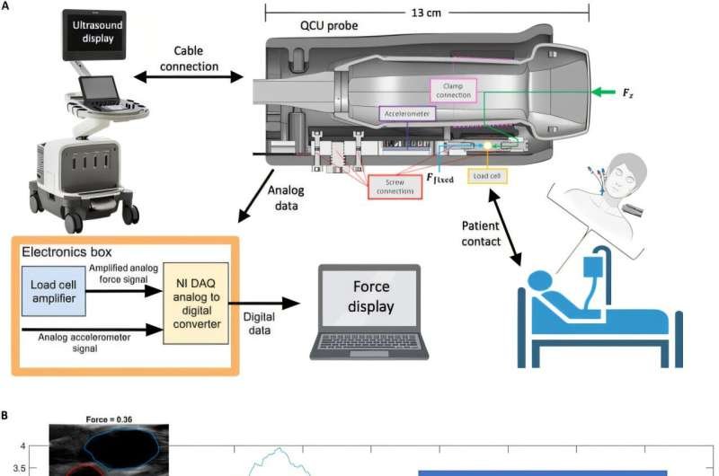
The study enrolled 11 patients from MGH’s cardiac intensive care unit (CICU), all of whom had indwelling central venous catheters as part of their standard care. Researchers meticulously captured QCU data, including short-axis, cross-sectional ultrasound images of the internal jugular vein (IJV), alongside simultaneous measurements of the force applied to the skin’s surface.
These noninvasive QCU measurements were then rigorously compared to the gold-standard invasive CVP data obtained directly from the patients’ central venous catheters. To further contextualize the findings, jugular venous pulsation height (JVP), a conventional noninvasive clinical measure, was also recorded in a subset of patients for comparative analysis.
The findings revealed a compelling correlation between the collapse force (CF)—defined as the force required to fully occlude the IJV short-axis cross-section—and invasive CVP measurements. Through linear regression analysis, the researchers achieved an impressive r² correlation coefficient of 0.82, with a mean absolute error of just 1.08 mmHg.
When accounting for hydrostatic pressure offset (HO), the correlation marginally increased to 0.83. In stark contrast, JVP measurements demonstrated a weaker correlation with invasive CVP, yielding an r² of 0.45 and a mean absolute error of 1.39 mmHg.
The ability to noninvasively and accurately estimate CVP carries profound clinical implications. CVP is a critical parameter in the management of patients with heart failure, sepsis, and other circulatory disorders, directly informing therapeutic interventions such as adjusting vasopressor medications or administering diuretics.
While traditional invasive methods offer accuracy, they are not without risks, including potential complications, and are resource-intensive. Noninvasive alternatives like JVP, on the other hand, are limited by inter-operator variability and dependence on patient positioning.
QCU stands out as a game-changing solution, providing reliable CVP estimates without the need for invasive procedures. This technology has the potential to streamline clinical workflows, reduce patient discomfort, and enable more frequent monitoring, particularly in resource-constrained environments.
This research was conducted by a collaborative team from the Massachusetts Institute of Technology (MIT) and Massachusetts General Hospital (MGH), Harvard Medical School. The team comprised Alex T. Jaffe from the Department of Electrical Engineering and Computer Science at MIT, Roger Pallarès-López and Jeffrey K. Raines from the Department of Mechanical Engineering at MIT, and Aaron D. Aguirre from the Cardiology Division at MGH, Harvard Medical School. The study was spearheaded by Brian W. Anthony, also from the Department of Mechanical Engineering at MIT.
More information:
Alex T. Jaffe et al, Noninvasive Quantitative Compression Ultrasound Central Venous Pressure: A Clinical Pilot Study, BME Frontiers (2025). DOI: 10.34133/bmef.0115
Provided by
BMEF (BME Frontiers)
Citation:
Noninvasive ultrasound method for measuring central venous pressure validated in clinical pilot study (2025, May 23)
retrieved 25 May 2025
from https://medicalxpress.com/news/2025-05-noninvasive-ultrasound-method-central-venous.html
This document is subject to copyright. Apart from any fair dealing for the purpose of private study or research, no
part may be reproduced without the written permission. The content is provided for information purposes only.